Home

strihať zobudiť sa čip retinal nerv fiber laser tomography ovládnuť únos vyrezávať

Optic Nerve and Retinal Nerve Fiber Imaging - EyeWiki

Scanning Laser Polarimetry Reveals Status of RNFL Integrity in Eyes with Optic  Nerve Head Swelling by OCT
Scanning Laser Polarimetry Reveals Status of RNFL Integrity in Eyes with Optic Nerve Head Swelling by OCT

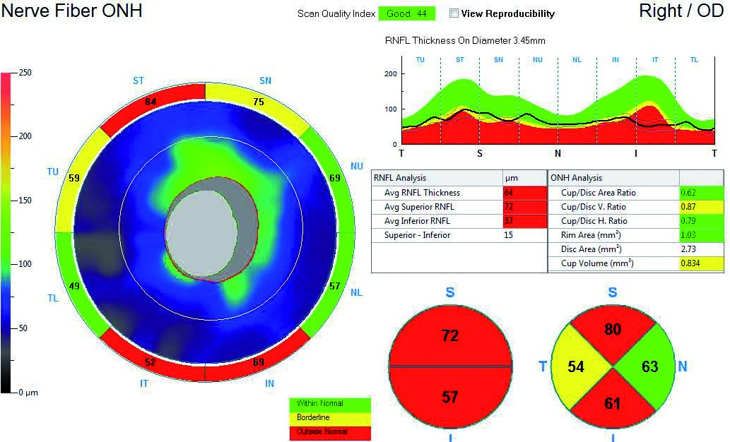
RNFL analysis with spectral-domain optical coherence tomography - American  Academy of Ophthalmology
RNFL analysis with spectral-domain optical coherence tomography - American Academy of Ophthalmology

Optical coherence tomography in multiple sclerosis: a systematic review and  meta-analysis - The Lancet Neurology
Optical coherence tomography in multiple sclerosis: a systematic review and meta-analysis - The Lancet Neurology

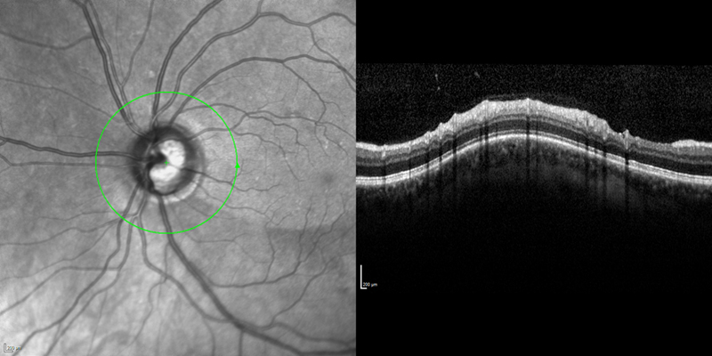
Optical coherence tomography of the retinal nerve fiber layer (RNFL)... |  Download Scientific Diagram
Optical coherence tomography of the retinal nerve fiber layer (RNFL)... | Download Scientific Diagram

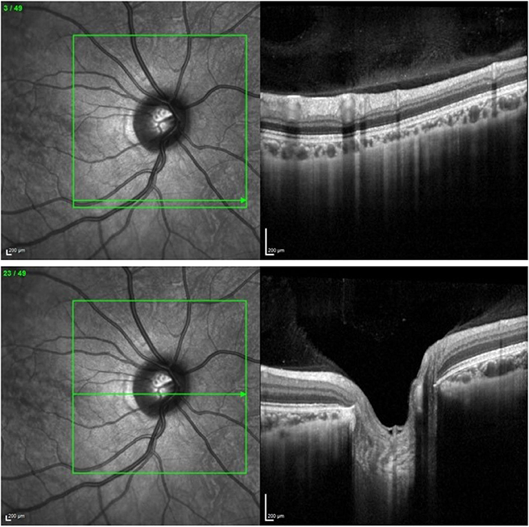
Optical Coherence Tomography of The Optic Disc | EYE DAY CLINIC

The application of optical coherence tomography in neurologic diseases |  Neurology Clinical Practice
The application of optical coherence tomography in neurologic diseases | Neurology Clinical Practice

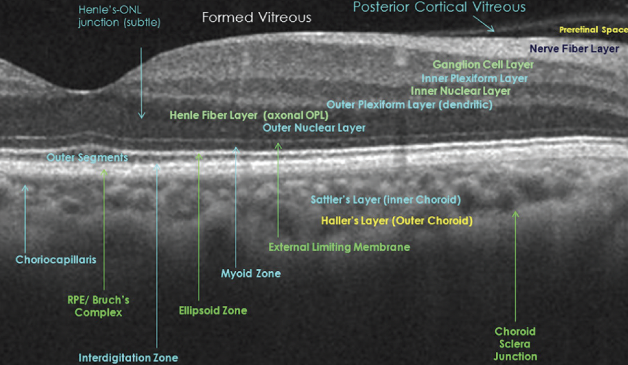
3 Optical Coherence Tomography of the Optic Nerve | Ento Key

Optical Coherence Tomography & Neurology - Practical Neurology

What OCT Tells Us About Progression

Optical coherence tomography/scanning laser ophthalmoscopy (OCT/SLO) of...  | Download Scientific Diagram
Optical coherence tomography/scanning laser ophthalmoscopy (OCT/SLO) of... | Download Scientific Diagram

A 3D model to evaluate retinal nerve fiber layer thickness deviations  caused by the displacement of optical coherence tomography circular scans  in cynomolgus monkeys (Macaca fascicularis) | PLOS ONE
A 3D model to evaluate retinal nerve fiber layer thickness deviations caused by the displacement of optical coherence tomography circular scans in cynomolgus monkeys (Macaca fascicularis) | PLOS ONE
Frontiers | Virtual Reality Improves Clinical Assessment of the Optic Nerve

Examinations of the optic nerve head

Retinal Nerve Fiber Layer - an overview | ScienceDirect Topics

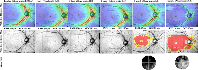
Traumatic optic neuropathy-associated progressive thinning of the retinal  nerve fiber layer and ganglion cell complex: two case reports | BMC  Ophthalmology | Full Text
Traumatic optic neuropathy-associated progressive thinning of the retinal nerve fiber layer and ganglion cell complex: two case reports | BMC Ophthalmology | Full Text

Figure 3 from Evaluating the optic nerve and retinal nerve fibre layer: the  roles of Heidelberg retina tomography, scanning laser polarimetry and  optical coherence tomography. | Semantic Scholar
Figure 3 from Evaluating the optic nerve and retinal nerve fibre layer: the roles of Heidelberg retina tomography, scanning laser polarimetry and optical coherence tomography. | Semantic Scholar

Comparison of Optical Coherence Tomography Measurement Reproducibility  between Children and Adults | PLOS ONE
Comparison of Optical Coherence Tomography Measurement Reproducibility between Children and Adults | PLOS ONE
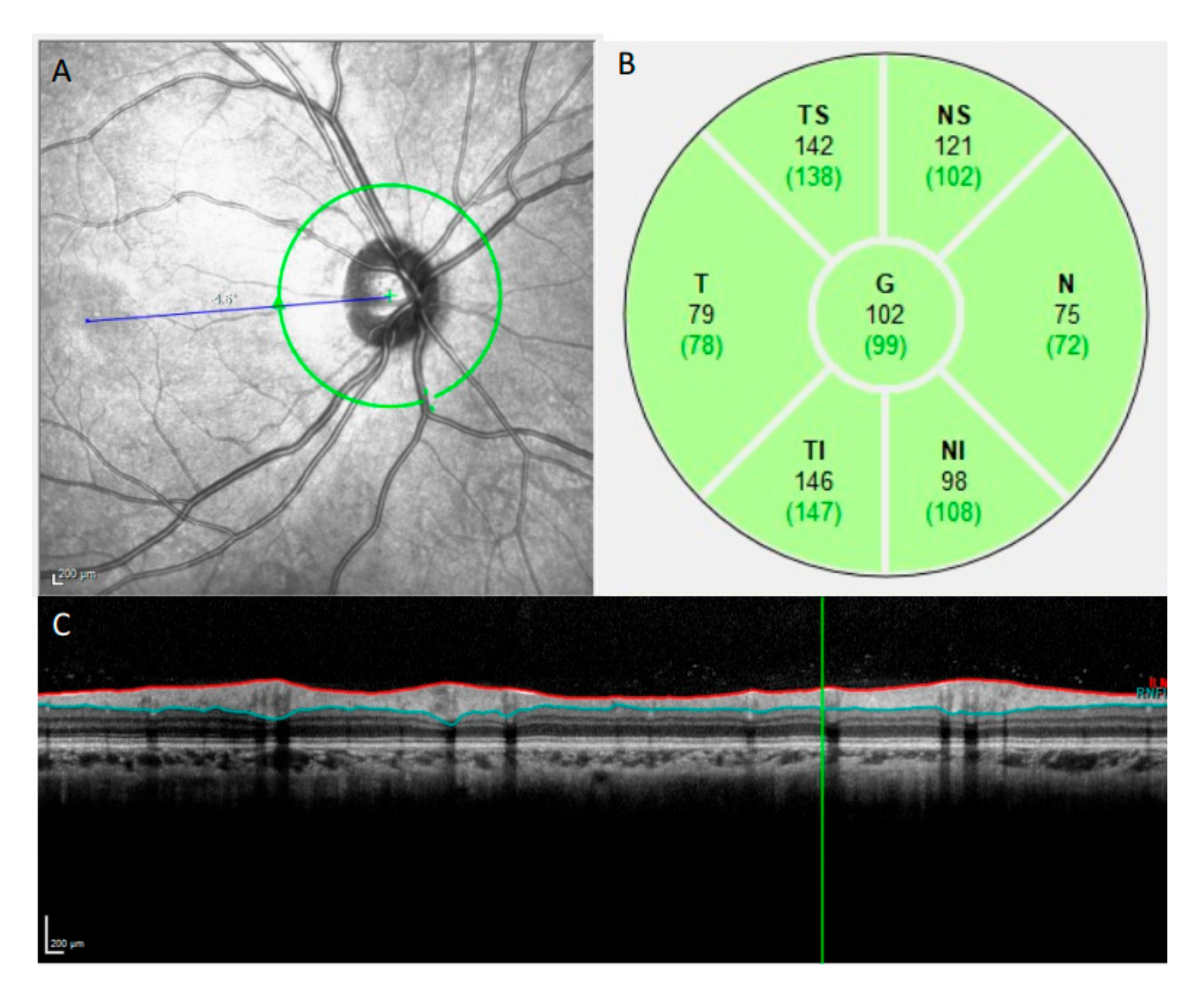
Nerve Fiber Analysis - Glaucoma Associates of Texas

Diagnostics | Free Full-Text | Thicker Retinal Nerve Fiber Layer with Age  among Schoolchildren: The Hong Kong Children Eye Study
Diagnostics | Free Full-Text | Thicker Retinal Nerve Fiber Layer with Age among Schoolchildren: The Hong Kong Children Eye Study

View Image
View Image

Visible light optical coherence tomography of peripapillary retinal nerve  fiber layer reflectivity in glaucoma | medRxiv
Visible light optical coherence tomography of peripapillary retinal nerve fiber layer reflectivity in glaucoma | medRxiv
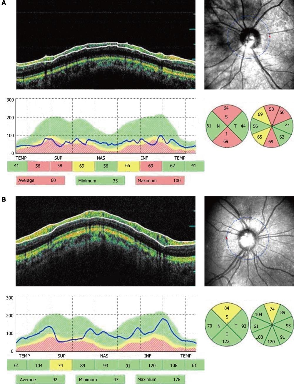
Changes in peripapillary retinal nerve fiber layer thickness in patients  with primary open-angle glaucoma after deep sclerectomy
Changes in peripapillary retinal nerve fiber layer thickness in patients with primary open-angle glaucoma after deep sclerectomy

Figure 1 | British Journal of Ophthalmology

View of Reproducibility of Retinal Nerve Fiber Layer and Macular Thickness  Measurements Using Spectral Domain Optical Coherence Tomography | Galician  Medical Journal
View of Reproducibility of Retinal Nerve Fiber Layer and Macular Thickness Measurements Using Spectral Domain Optical Coherence Tomography | Galician Medical Journal

Original and delineated spectral-domain optical coherence tomography... |  Download Scientific Diagram
Original and delineated spectral-domain optical coherence tomography... | Download Scientific Diagram